| Banana Diseases | ||||||||||||||||||||||||||||||||||||||||||||||||||||||||||||||||||
|---|---|---|---|---|---|---|---|---|---|---|---|---|---|---|---|---|---|---|---|---|---|---|---|---|---|---|---|---|---|---|---|---|---|---|---|---|---|---|---|---|---|---|---|---|---|---|---|---|---|---|---|---|---|---|---|---|---|---|---|---|---|---|---|---|---|---|
| Back
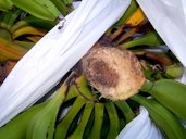
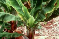
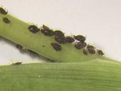
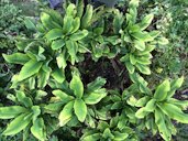
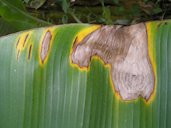
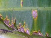
to Banana Page  Fig. 1  Wilt symptom, Panama disease of banana 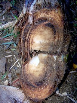 Fig. 2  Internal necrosis of banana pseudostem, a symptom of Panama wilt disease 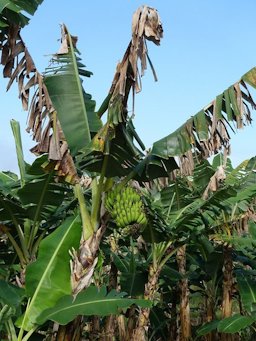 Fig. 10  Black leaf streak (Black Sigatoka), pathogen: Mycosphaerella fijiensis (fungus) 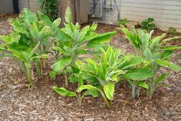 Fig. 17  Typical symptoms of banana bunchy top caused by the banana bunchy top virus (BBTV) 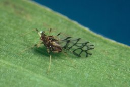 Fig. 18  Winged (alate) banana aphid (Pentalonia nigronervosa), vector of banana bunchy top virus 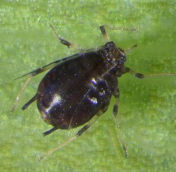 Fig. 27  Adult banana aphid (Pentalonia nigronervosa) 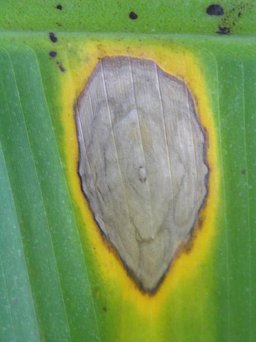 Fig. 28  Cordana leaf spot of banana, caused by Cordana musae
|
Panama Disease
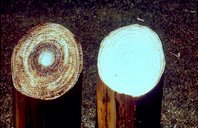
(Fusarium wilt) Fusarium oxysporum f. sp. cubense Panama disease is of worldwide importance and is caused by the soil borne fungus Fusarium oxysporum f. sp. cubense. There are four known races of the disease, three of which attack one or more banana cultivars (race 1,2 and 4). Symptoms of the disease do not appear on young suckers. On mature plants symptoms include progressive yellowing and eventual death from older to younger leaves, so that only the youngest emerging leaf may remain; brown and black discoloration and slimy appearance of the water conducting vascular system (it may give off a bad odor as well); and death of the plant. At present there is no chemical control available. The only effective control measures are planting in land not infested with the fungus, the use of disease-free propagation material, and the planting of cultivars with resistance to the disease. Plantains are resistant to the fungus. 1
Fig. 3. Panama wilt (historical) Fig. 4,5,6. Panama wilt disease of banana Fig. 7. Panama disease of banana: rotten peduncle Fig. 8. Internal necrosis of banana pseudostem, a symptom of Panama wilt disease Fig. 9. Panama wilt of banana (left); necrotic tissue inside pseudostem; healthy pseudostem (right)
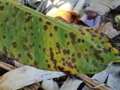
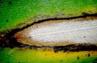
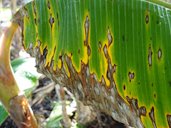
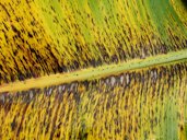
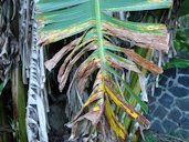
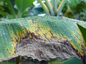
Further Reading Fusarium wilt or Panama disease Historic Overview, Food and Agriculture Organization of the United Nations pdf Sigatoka (Yellow Sigatoka and Black Sigatoka) Pathogen: Mycosphaerella fijiensis /Pseudocercospora fijiensis Black sigatoka and yellow sigatoka are of worldwide importance; in general, where the two diseases are found, Black sigatoka dominates as the most severe disease causing fungus. Black sigatoka is an important leaf disease in Florida. Yellow sigatoka is caused by the fungus Mycosphaerella musicola and black sigatoka M. fijiensis. Symptoms of yellow sigatoka begin as pale green flecks that become brown with yellow haloes. As the disease progresses infected areas coalesce forming large areas of dead leaf tissue. Black sigatoka begins as minute reddish-brown flecks on the lower leaf surface but as the infection progresses dark flecks may be seen on the upper leaf surface as well. As the disease progresses the dark areas with yellow haloes surrounding dead leaf tissue coalesce until the entire leaf is killed. Warm temperatures, high humidity, and frequent rainfall are ideal for disease development. Sigatoka does not kill the plant but causes premature defoliation which results in reduced crop yield. 1 Banana cultivars differ in their susceptibility to sigatoka with the Cavendish group (AAA) and 'Pome' (AAB) bananas being highly susceptible. 'Sucrier' (AA), 'Bluggoe' (ABB), and 'Silk' (AAB) are of intermediate susceptibility, while 'Mysore' is only slightly susceptible. Fungicides are available for control but may not be necessary for banana plants in the home landscape. For more information, please contact your local Cooperative Extension Agent. 1
Fig. 11. The streaks are absent on the leaves of young plants. Instead, circular leaf spots form Fig. 12. Sigatoka fungal disease. Black leaf streak lesion on banana leaf Fig. 13. Black leaf streak (Black Sigatoka), pathogen: M. fijiensis (fungus) Fig. 14,15. Black leaf streak aka Black Sigatoka Fig. 16. In areas with high rainfall, necrotic banana leaves turn very dark in color as the dead tissues becomes waterlogged; hundreds of smaller black leaf streak lesions coalesce to form large, blighted areas on banana leaves
Further Reading Black Leaf Streak of Banana, University of Hawai'i at Mānoa pdf Banana bunchy top virus (BBTV) in Hawai'i Vector: Black banana aphid, Pentalonia nigronervosa Banana bunchy top virus (BBTV) is one of the most serious diseases of banana. Once established, it is extremely difficult to eradicate or manage. BBTV is widespread in Southeast Asia, the Philippines, Taiwan, most of the South Pacific islands, and parts of India and Africa. BBTV does not occur in Central or South America. Symptoms: reduced internode distance, chlorotic leaf margins, erect leaf architecture, plant stunting, green J-hooks, mottled petioles and leaf sheaths, Morse code in leaf vines.
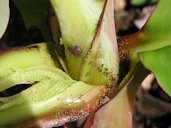
Fig. 19. Petiole mottling, a symptom of banana bunchy top Fig. 20. Banana bunchy top disease: green J-hooks and Morse code in leaf venation Fig. 21. Mottled banana inflorescence infected with the banana bunchy top virus Fig. 22. Symptoms of banana bunchy top disease in older banana plant Fig. 23. Banana bunchy top disease symptoms on a young 'Williams' hybrid banana plant Fig. 24. Colony of banana aphids (P. nigronervosa), vector of banana bunchy top virus; they often hide under the banana leaf sheath Fig. 25. Colony of banana aphids (P. nigronervosa), vector of banana bunchy top virus Fig. 26. These plants arose from the pseudostem of a central, infected mother plant that was cut down.
Further Reading Bunchy Top Virus, University of Hawai'i at Mānoa pdf Banana bunchy top virus and its vector Pentalonia nigronervosa, Florida Department of Agriculture and Consumer Services pdf Banana Bunchy Top: Detailed Signs and Symptoms, University of Hawai'i at Mānoa pdf Cordana Leaf Spot caused by the pathogen Cordana musae
Fig. 29,30. Cordana leaf spot of banana, caused by Cordana musae Other Banana Diseases
Postharvest Rots of Banana, University of Hawai'i at Mānoa pdf |
|||||||||||||||||||||||||||||||||||||||||||||||||||||||||||||||||
| Bibliography 1 Crane, Jonathan H., and Carlos F. Balerdi. "Bananas Growing in the Home Landscape." Hotcicultural Service Dept., UF/IFAS,Extension, HS10, Original pub. Oct. 1971 as FC-10, Revised Jan. 1998, Dec. 2005, Oct. 2008, and Nov. 2016, Reviewed Dec. 2019, AskIFAS, edis.ifas.ufl.edu/mg040. Accessed 24 Mar. 2017, 3 Aug. 2020. Videos v 1,2,4,5,6,7,8,9 Crane, Johathan H., and Ian Maguire. "Banana Video Series." University of Florida, IFAS/TREC. v 3 Nelson, Scot C., and L. Richardson. "Banana Bunchy Top in Hawaii." University of Hawai'i at Mānoa, Written, directed and produced by Scot C. Nelson, Filmed and edited by Lynn Richardson, Narrated by John Burnett, Music by Boone Morrison, Sept. 2006, CTAHR, www.ctahr.hawaii.edu/banana/video.asp. Accessed 7 May 2018. Photographs Fig. 1 Nelson, Scot C. "Wilt symptom, Panama disease of banana." Flickr, 2004, (CC BY-SA 2.0), www.flickr.com. Accessed 20 March 2017. Fig. 2,8 Nelson, Scot C. "Internal necrosis of banana pseudostem, a symptom of Panama wilt disease." Flickr, 2006, (CC BY-SA 2.0), www.flickr.com. Accessed 20 March 2017. Fig. 3 Trujillo, E. E. "Banana: Panama wilt (historical)." Flickr, 2012, (CC BY-SA 2.0), www.flickr.com. Accessed 20 March 2017. Fig. 4,5,6 Nelson, Scot C. "Panama wilt disease of banana." Flickr, 2005, (CC BY-SA 2.0), www.flickr.com. Accessed 20 March 2017. Fig. 7 Nelson, Scot C. "Panama disease of banana: rotten peduncle." Flickr, 2006, (CC BY-SA 2.0), www.flickr.com. Accessed 20 March 2017. Fig. 9 Nelson, Scot C. "Panama wilt of banana (left). Necrotic tissue inside pseudostem. Healthy pseudostem (right)." Flickr, 2014, (CC BY-SA 2.0), www.flickr.com. Accessed 20 March 2017. Fig. 10,13,16 Nelson, Scot C. "Banana: Black leaf streak (Black sigatoka). Pathogen: Mycosphaerella fijiensis (fungus)." Flickr, 2016, (CC BY-SA 2.0), www.flickr.com. Accessed 20 March 2017. Fig. 11 Nelson, Scot C. "The streaks are absent on the leaves of young plants. Instead, circular leaf spots form." Flickr, 2016, (CC BY-SA 2.0), www.flickr.com. Accessed 20 March 2017. Fig. 12 Nelson, Scot C. "Sigatoka fungal. Black leaf streak lesion on banana leaf." Flickr, 2007, (CC BY-SA 2.0), www.flickr.com. Accessed 20 March 2017. Fig. 14,15 Nelson, Scot C. "Banana: Black leaf streak aka Black Sigatoka." Flickr, 2015, (CC BY-SA 2.0), www.flickr.com. Accessed 20 March 2017. Fig. 17 Nelson, Scot C. "Typical symptoms of banana bunchy top caused by the banana bunchy top virus (BBTV)." Flickr, 2012, (CC BY-SA 2.0), www.flickr.com. Accessed 17 March 2017. Fig. 18 Nelson, Scot C. "Winged (alate) banana aphid (Pentalonia nigronervosa), vector of banana bunchy top virus (BBTV)." Flickr, 2011, (CC BY-SA 2.0), www.flickr.com. Accessed 17 March 2017. Fig. 19 Nelson, Scot C. "Petiole mottling, a symptom of banana bunchy top." Flickr, 2008, (CC BY-SA 2.0), www.flickr.com. Accessed 17 March 2017. Fig. 20 Nelson, Scot C. "Banana bunchy top disease: Green J-hooks and Morse code in leaf venation." Flickr, 2003, (CC BY-SA 2.0), www.flickr.com. Accessed 17 March 2017. Fig. 21 Nelson, Scot C. "Mottled banana inflorescence infected with the banana bunchy top virus (BBTV)." Flickr, 2011, (CC BY-SA 2.0), www.flickr.com. Accessed 17 March 2017. Fig. 22 Nelson, Scot C. "Symptoms of banana bunchy top disease in older banana plant." Flickr, 2011, (CC BY-SA 2.0), www.flickr.com. Accessed 17 March 2017. Fig. 23 Nelson, Scot C. "Banana bunchy top disease symptoms on a young 'Williams' hybrid banana plant." Flickr, 2011, (CC BY-SA 2.0), www.flickr.com. Accessed 17 March 2017. Fig. 24 Nelson, Scot C. "Colony of banana aphids (Pentalonia nigronervosa), vector of banana bunchy top virus (BBTV). They often hide under the banana leaf sheath." Flickr, 2004, (CC BY-SA 2.0), www.flickr.com. Accessed 17 March 2017. Fig. 25 Nelson, Scot C. "Colony of banana aphids (Pentalonia nigronervosa), vector of banana bunchy top virus (BBTV)." Flickr, 2004, (CC BY-SA 2.0), www.flickr.com. Accessed 17 March 2017. Fig. 26 Nelson, Scot C. "These plants arose from the pseudostem of a central, infected mother plant that was cut down." Flickr, 2014, (CC BY-SA 2.0), www.flickr.com. Accessed 20 March 2017. Fig. 27 Nelson, Scot C. "Adult banana aphid (Pentalonia nigronervosa)." Flickr, 2004, (CC BY-SA 2.0), www.flickr.com. Accessed 20 March 2017. Fig. 28 Nelson, Scot C. "Cordana leaf spot of banana, caused by Cordana musae." Flickr, Sept. 14, 2006, Public Domain, www.flickr.com/photos/scotnelson/5693515317/in/album-72157645335860420/. Accessed 24 Apr. 2018. Fig. 29 Nelson, Scot C. "Cordana leaf spot of banana, caused by Cordana musae." Flickr, Sept. 14, 2006, Public Domain, www.flickr.com. Accessed 24 Apr. 2018. Fig. 30 Nelson, Scot C. "Cordana leaf spot of banana, caused by Cordana musae." Flickr, Sept. 14, 2006, Public Domain, www.flickr.com/5693515391/in/album-72157645335860420/. Accessed 24 Apr. 2018. Published 17 Apr. 2014 LR. Last update 8 Mar. 2023 LR |
||||||||||||||||||||||||||||||||||||||||||||||||||||||||||||||||||


